Visual atrophy
Visual atrophy
Visual atrophy

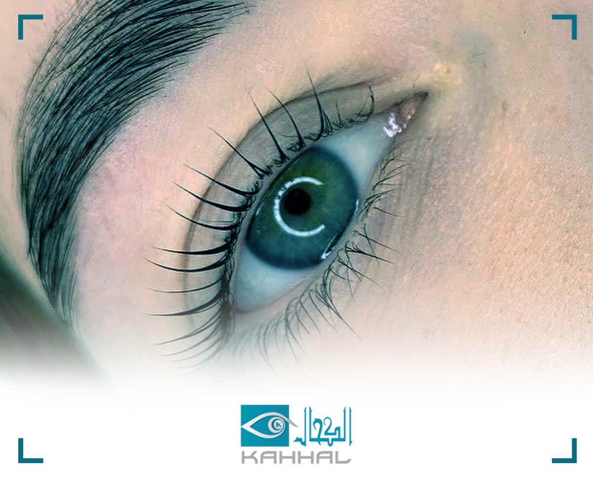
Optic atrophy (or optic disc atrophy) is a definitive, viable condition of diseases that damage the mental cell (Ganglion cell) and the optic nerve fibers. Symptoms of visual impairment: A feeling of decreased color vision and a decrease in image clarity. - A decrease in vision (not always), a defect in color vision and a relative dysfunction of the pupils in cases of unilateralism or asymmetry, disc view - in the fundus of eye examination, pallor and sagging of the nerve fibers appear. A decrease in the field of vision. Causes and risk factors of visual atrophy: Some of the causative factors lead to atrophy on both sides and others to atrophy on one side only. Eyes: Uveitis, Glaucoma, Pigmentosa retinitis. Orbital nerve: a tumor in the nerve (glioma), a tumor in the covering of the nerve (meningioma), a tumor in the orbital that presses on the optic nerve. Brain: Intracranial pressure (intracranial pressure) is high for unknown reasons, a tumor presses on a nerve directly, or increases intracranial pressure, an acute aneurysm pressing on a nerve. Myelin infections in the nerve sheaths and in the nerve itself. Poisoning is possible from drugs, for example: digitalis (a genus of heart-strengthening medicinal herbs - Digitalis), isoniazid (Isonizid), or poisoning from some substances: such as methanol. Hereditary disorders that appear in childhood, such as Dominant optic atrophy, or other disorders that appear, mainly, in men in the second or third decade, Optic neuropathy leber. Injury to the face or head. Disturbance in the flow and supply of blood. Diagnosing visual atrophy: The patient’s narration about the medical history and the sequence of events as they appeared has a very important importance in the diagnosis process, as it is directed towards the correct and accurate diagnosis, for example: Is the conversation about a sudden and severe emergence of the disease? Or is it a slow onset that develops gradually? Is there any pain? Has there been an accident that resulted in a head / face injury? Does the patient take any medications? Does he smoke a lot? Does he drink alcohol? ... and others. According to the results of the examinations and in the continuation of the clarification process, the patient is directed to perform additional assistive examinations, blood tests, x-ray imaging (x-ray) and compyted tomography (ct) magnetic resonance imaging (MRI). , Nerve and retinal conduction testing, visual evoked potential -VED and electroretinography - ERG.